Pulmonary Eosinophilia Indicates Onset of Allergic Bronchopulmonary Aspergillosis
By LabMedica International staff writers
Posted on 13 Dec 2021
Allergic bronchopulmonary aspergillosis (ABPA) is a pulmonary disorder caused by type 2 immune responses to Aspergillus. This pathology usually develops after the onset of bronchial asthma (BA) or cystic fibrosis. Rates differ among countries, but appear to be increasing around the world with the increase of BA.Posted on 13 Dec 2021
Several diagnostic criteria for ABPA have been proposed, such as a history of BA or cystic fibrosis, characteristic radiographic pulmonary opacities including central dilatation of the bronchus, elevated immunoglobulin (Ig)E levels, and hypersensitivity reaction to Aspergillus including elevated IgE antibodies against Aspergillus fumigatus and/or IgG antibodies for Aspergillus. ABPA and chronic eosinophilic pneumonia (CEP) both display peripheral eosinophilia as well as pulmonary infiltration, together described as pulmonary eosinophilia, and differentiation is sometimes problematic.
")
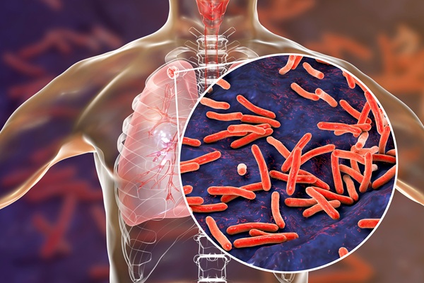
Image: Histopathology of Allergic bronchopulmonary aspergillosis. Grocott\'s Methenamine Silver Stain shows branching and helps to confirm that the black structures are indeed fungal hyphae (Photo courtesy of Doctorlib)
Pulmonologists at the Osaka Toneyama Medical Center (Osaka, Japan) performed a retrospective, single-center study that included patients that fulfilled the diagnostic criteria for ABPA, which included a history of BA or cystic fibrosis and both of the following: positive immediate cutaneous hypersensitivity to Aspergillus antigen or specific IgE to A. fumigatus, and elevated total IgE level in serum > 1000 IU/mL. Also the presence of precipitating or IgG antibodies against A. fumigatus in serum; pulmonary opacities consistent with ABPA on radiographic imaging; and total eosinophil count > 500 cells/µL in steroid-naïve patients were considered.
Patients were assigned to either the eosinophilic pneumonia (EP) group or Non-EP group, defined according to findings on high-resolution computed tomography (HRCT). The EP group included patients with HRCT findings compatible with CEP; i.e., the presence of peripheral consolidation (p-consolidation) or ground-glass opacities (GGO), with no evidence of high-attenuation mucus. The Non-EP group comprised the remaining patients, who showed classical findings of ABPA such as mucoid impaction. Differences between the groups were analyzed.
The investigators reported that baseline characteristics, frequency of a history of CEP (EP, 50% versus Non-EP, 26%) and tentative diagnosis of CEP before diagnosis of ABPA (67% versus 16%) did not differ significantly between groups. Although elevated absolute eosinophil count and Aspergillus-specific immunoglobulin E titers did not differ significantly between groups, the Non-EP group showed a strong positive correlation between these values (R = 0.7878). All patients showed peripheral eosinophilia (median, 1,540 cells/μL), elevated level of IgE (median, 2,802 IU/mL) and positive reactions for Aspergillus-specific IgE (median, 20.7 IU/mL).
The Non-EP group displayed significantly higher levels of the fungal marker beta-D glucan (median, 11.7 pg/mL; interquartile range, 6.7–18.4 pg/mL) than the EP group (median, 6.6 pg/mL; interquartile range, 5.2–9.3 pg/ml). Both groups exhibited frequent recurrence of shadows on X-rays but no cases in the EP group had progressed to the Non-EP group at the time of relapse.
The authors concluded that the ABPA subgroup with imaging findings resembling CEP experienced frequent recurrences, as in typical ABPA. In pulmonary eosinophilia, even if there are no shadows indicating apparent mucous change, the Aspergillus-specific immunoglobulin E level is important in obtaining an accurate diagnosis and in the selection of appropriate therapies for this type of ABPA. The study was published on November 18, 2021 in the journal Allergy, Asthma & Clinical Immunology.
Related Links:
Osaka Toneyama Medical Center
.jpg)
Gold Member
Clinical Chemistry Assay
Sorbitol Dehydrogenase (SDH)


Chromogenic Culture System
InTray™ COLOREX™ ECC

Electrolyte Analyzer
BKE-B