Plasma Amyloid-β 42/40 Assays Compared in Alzheimer Disease
By LabMedica International staff writers
Posted on 14 Oct 2021
Blood-based tests for brain amyloid-β (Aβ) pathology are needed for widespread implementation of Alzheimer disease (AD) biomarkers in clinical care and to facilitate patient screening and monitoring of treatment responses in clinical trials.Posted on 14 Oct 2021
Reliable measurements of Aβ in blood proved challenging until the development of advanced mass spectrometry and immunodetection methods. Recent articles have suggested that Aβ42/40 quantified using ultrasensitive and fully automated immunoassay platforms could predict Aβ-PET status (especially when combined with APOE genotype) with accuracy approaching that of MS-based Aβ42/40 measures.
")
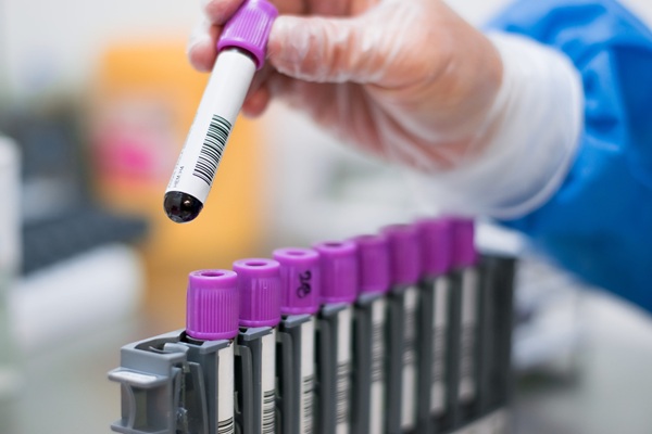
Image: Simoa is an ultra-sensitive immunoassay technology that allow detection of proteins and nucleic acids at lowest possible levels (Photo courtesy of Quanterix)
An international team led by Clinical Neuroscientists at Lund University (Lund, Sweden) compared the performance of plasma Aβ42/40 measured using eight different Aβ assays when detecting abnormal brain Aβ status in patients with early AD. The study included 182 cognitively unimpaired participants and 104 patients with mild cognitive impairment who were enrolled at three different hospitals in Sweden and underwent Aβ positron emission tomography (PET) imaging and cerebrospinal fluid (CSF) and plasma collection from 2010 to 2014.
Plasma Aβ42/40 was measured using an immunoprecipitation-coupled mass spectrometry developed at Washington University School of Medicine (IP-MS-WashU, St Louis, MO, USA), antibody-free liquid chromatography MS developed by Araclon (LC-MS-Arc, Zaragoza, Spain), and diverse immunoassays. Plasma Aβ42/40 was also measured using an IP-MS–based method from Shimadzu (Kyoto, Japan) in 200 participants (IP-MS-Shim) and an IP-MS–based method from the University of Gothenburg (IP-MS-UGOT, Gothenburg, Sweden) and another immunoassay, the N4PE Simoa immunoassay (IA-Quan, Quanterix Billerica, MA, USA) among 227 participants.
When the team identified participants with abnormal CSF Aβ42/40 in the whole cohort, plasma IP-MS-WashU Aβ42/40 showed significantly higher accuracy than LC-MS-Arc Aβ42/40, and some immunoassays. Plasma IP-MS-WashU Aβ42/40 performed significantly better than IP-MS-UGOT Aβ42/40 and IA-Quan Aβ42/40, while there was no difference in the AUCs between IP-MS-WashU Aβ42/40 and IP-MS-Shim Aβ42/40) in the two sub-cohorts where these biomarkers were available. Plasma IPMS-WashU Aβ42/40 and IP-MS-Shim Aβ42/40 showed highest coefficients for correlations with CSF Aβ42/40.
The authors concluded that the results from two independent cohorts indicate that certain MS-based methods performed better than most of the immunoassays for plasma Aβ42/40 when detecting brain Aβ pathology. The study was published on September 20, 2021 in the journal JAMA Neurology.
Related Links:
Lund University
Washington University School of Medicine
Araclon
University of Gothenburg
Quanterix

Gold Member
Automatic Hematology Analyzer
CF9600


Automatic CLIA Analyzer
Shine i6000

Japanese Encephalitis Test
Japanese Encephalitis Virus Real Time PCR Kit