Fluorimetric Assay Quantifies Galactocerebrosidase Activity in Dried Blood Spots
By LabMedica International staff writers
Posted on 29 Oct 2019
The lysosomal enzyme galactocerebrosidase hydrolyzes glycosidic bonds of several glycosphingolipids, including galactose from galactosylsphingosine (psychosine), and is essential to prevent the toxic accumulation of psychosine in the body.Posted on 29 Oct 2019
Decreased galactocerebrosidase (GALC) enzyme activity is causative for Krabbe disease, a lysosomal storage disorder with devastating neurodegenerative consequences. Quantitative fluorimetric assays for GALC activity in isolated blood and skin cells have been described, but not for dried blood spots specimens (DBS).
.")
Image: The Synergy HTX multi-mode microplate reader is a compact, affordable system for 6- to 384-well microplates and Take3 Micro-Volume Plates (Photo courtesy of BioTek).
A team of scientists from the commercial company Baebies, Inc (Durham, NC, USA) and Duke University (Durham, NC, USA) developed a rapid, microtiter plate fluorimetric assay for measuring GALC enzyme activity in DBS specimens using a novel substrate: β-galactose conjugated with a fluorogenic derivative of 6-hexadecanoyl-4-methylumbelliferone with a hydrophobic group.
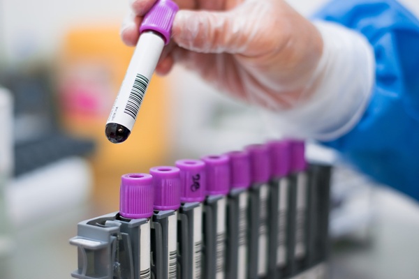
Samples were obtained as individual punches (3.2 mm diameter) from DBS cards of presumed normal newborns. Archived, deidentified DBS from 10 affected Krabbe disease patients were obtained from the Legacy of Angels Foundation. To extract galactocerebrosidase enzyme from the DBS samples, one punch (3.2 mm) from each DBS was placed in individual wells of a clear, round-bottom, 96-well microtiter plate.
Sample extraction solution (100 μL) was added to each sample well; the plate was covered with a clear adhesive sealer to prevent evaporation and then incubated on a plate-shaker (600 rpm) at room temperature (RT) for 30 minutes. Enzyme activity was determined by adding 10 μL of DBS extract to 10 μL of the GALC substrate solution, which was varied. The fluorescence of the plate, measured as relative fluorescence units (RFU), was read in a Synergy HTX microtiter plate reader with 400 ±15 nm excitation and 485 ±20 nm emission filters.
The GALC assay was carefully optimized to ensure robust performance from the small amount of enzyme present in DBS and to minimize interference from β-galactosidase. The team found that the linear range of the fluorimetric GALC assay encompassed the entire range of samples tested. The activity in the presumed normal samples shows a wide range (0.39 – 15.6 μmol/L/hour) with a population mean of 2.108 μmol/L/hour. As expected, GALC activity in the affected samples is significantly lower than in the presumed normal samples.
The authors concluded that a fluorimetric assay for GALC enzyme activity measurement on dried blood spot specimens is feasible. Improvements to the assay including novel substrate design, increased substrate concentration and removal of sodium chloride maximize the specificity of the assay and minimize interference from β-galactosidase. The study was published on October 16, 2019, in the journal Practical Laboratory Medicine.
Related Links:
Baebies
Duke University

Gold Member
Automatic Hematology Analyzer
CF9600


Multi-Chamber Washer-Disinfector
WD 390

Japanese Encephalitis Test
Japanese Encephalitis Virus Real Time PCR Kit