Biomarkers Panel Used for Pleural and Peritoneal Effusions Diagnosis
By LabMedica International staff writers
Posted on 25 Sep 2019
The quantification of biomarkers in cavity fluids may aid in the etiological diagnosis and in patients' management. Laboratory investigation of cavity effusions involves the evaluation of biochemical, immunological, microbiological, molecular and cellular parameters.Posted on 25 Sep 2019
The broad spectrum of etiologic causes of pleural effusion (PE) and peritoneal effusion (PerE) justifies studies with biomarkers quantified in samples obtained through thoracentesis and/or paracentesis, procedures considered minimally invasive and at a low risk for patients.
.")
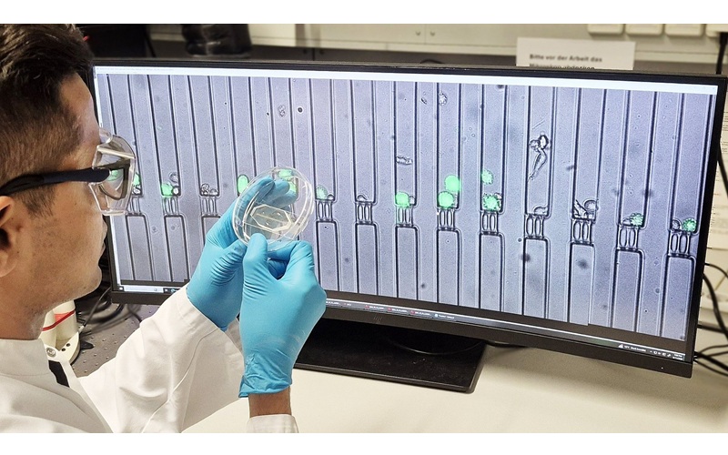
Image: The MAGPIX fluorescent-based analytical instrument (Photo courtesy of Luminex).
Scientists at the University of Sao Paulo (San Paulo, Brazil) evaluated Samples of pleural or peritoneal fluid from 120 patients submitted to thoracentesis or paracentesis for diagnostic investigation. Each sample was representative of one patient. The team evaluated the following biomarkers: carcinoembryonic antigen (CEA), vascular endothelial growth factor A (VEGF-A) programmed death-ligand 1 (PD-L1/B7-H1), neutrophil gelatinase-associated lipocalin (NGAL), triggering receptor expressed in myeloid cells type-1(TREM-1), and gamma-interferon (IFNγ).
Samples of PE or PerE were prepared for analysis in a 96-well plate utilizing a custom Luminex Human Magnetic Assay. CEA, VEGF-A, PD-L1, NGAL, TREM-1 and IFNγ were quantified using a MAGPIX analytical test instrument. The concentrations of Calprotectin were obtained by a ‘sandwich’ ELISA test with CALP human kit and analyzed on Victor X3 plate reader in a 450 nm low-pass absorbance. The assay used for adenosine desaminase (ADA) was the Quimiada Adenosina Deaminase kit, and a concentration above 30 U/L is presumptive of tuberculosis.
The team reported that for malignant effusion (ME) diagnosis, CEA and NGAL presented superior performance than VEGF-A, PD-L1 and CALP. A CEA-NGAL association showed good sensitivity (86.6%) and accuracy (79.2%). For non-tuberculous infectious effusion (NTBIE), NGAL presented the best performance with sensitivity (75.0%), specificity (62.0%) and accuracy (65.0%) higher than TREM-1 and CALP; however, when associated, although with good sensitivity, there was important decrease in specificity. For tuberculous pleural effusion (TPE), IFNγ-ADA presented excellent sensitivity (100%), specificity (87.6%), NPV (100%) and accuracies (~90%).
The authors concluded that a hybrid panel composed by the biomarkers CEA, NGAL, IFNγ and ADA seems to be useful in discriminating between ME and TPE etiology, both lymphocytic effusions. For non-tuberculous infectious effusion, the panel used did not demonstrate diagnostic advantages over the classic literature parameters. The study was published in the October 2019 issue of the journal Clinica Chimica Acta.
Related Links:
University of Sao Paulo

Gold Member
Quantitative POC Immunoassay Analyzer
EASY READER+


Multi-Chamber Washer-Disinfector
WD 390

HPV Molecular Test
BD Onclarity HPV Assay