Cause of NMO Illuminated by High-Tech Microscope
By LabMedica International staff writers
Posted on 10 May 2017
Neuromyelitis optica (NMO), also known as Devic's disease or Devic's syndrome, is a heterogeneous condition consisting of the simultaneous inflammation and demyelination of the optic nerve (optic neuritis) and the spinal cord (myelitis) and it can be monophasic or recurrent.Posted on 10 May 2017
Determining the spatial relationship of individual proteins in dense assemblies remains a challenge for super-resolution nanoscopy. A unique microscope capable of illuminating living cell structures in great detail has been used to find clues into how this destructive autoimmune disease works, setting the stage for more discoveries in the future.
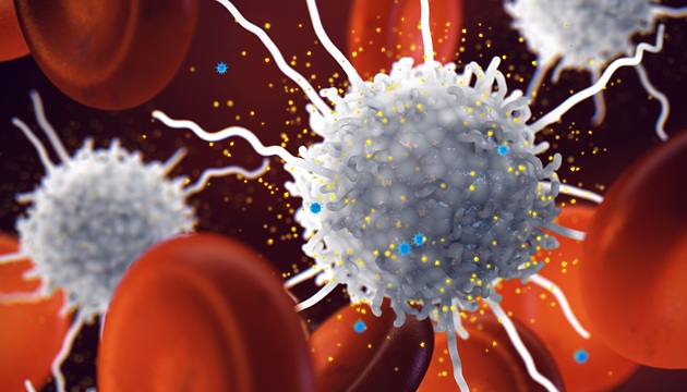
 that affects the optic nerves and spinal cord (Photo courtesy of Dr. William Pawluk).")
Image: Researchers used a custom STED microscope to determine the cause of NMO, an uncommon disease syndrome of the central nervous system (CNS) that affects the optic nerves and spinal cord (Photo courtesy of Dr. William Pawluk).
Biophysicists at the University of Colorado Anschutz Medical Campus used a custom Stimulated Emission Depletion (STED) microscope built at CU Anschutz; they were able to actually see clusters of antibodies atop astrocytes, the brain cell target of the autoimmune response in NMO. They imaged secondary antibody labeling of monoclonal aquaporin-4- immunoglobulin G (AQP4-IgGs) with differing epitope specificity bound to isolated tetramers (M1-AQP4) and large orthogonal arrays of AQP4 (M23-AQP4).
Imaging secondary antibodies bound to M1-AQP4 allowed the team to infer the size of individual AQP4-IgG binding events. This information was used to model the assembly of larger AQP4-IgG complexes on M23-AQP4 arrays. A scoring algorithm was generated from these models to characterize the spatial arrangement of bound AQP4-IgG antibodies, yielding multiple epitope-specific patterns of bound antibodies on M23-AQP4 arrays.
The authors concluded that their results delineate an approach to infer spatial relationships within protein arrays using stimulated emission depletion nanoscopy, offering insight into how information on single antibody fluorescence events can be used to extract information from dense protein assemblies under a biologic context. Jeffrey Bennett, MD, PhD, a professor and senior author of the study, said, “We discovered that we could see the natural clustering of antibodies on the surface of target cells. This could potentially correspond with their ability to damage the cells. We know that once antibody binds to the surface of the astrocyte, we are witnessing the first steps in the disease process.” The study was published on April 25, 2017, issue of the Biophysical Journal.
.jpg)
Gold Member
Clinical Chemistry Assay
Sorbitol Dehydrogenase (SDH)


Japanese Encephalitis Test
Japanese Encephalitis Virus Real Time PCR Kit

Hematology Consumables
Bioblood Devices