Increased Serum Cytokine Concentrations Reported in Guillain-Barré Syndrome
By LabMedica International staff writers
Posted on 05 Oct 2016
Guillain-Barré syndrome (GBS) is an acquired demyelinating peripheral neuropathy and it has been shown that macrophage activation contribute to the pathogenesis of GBS. Therefore macrophage-mediated factors could be the potential markers for disease diagnosis and status of GBS.Posted on 05 Oct 2016
The diagnosis of GBS is based on a combination of clinical, electrophysiological features as well as analysis of the cerebrospinal fluid (CSF). A biomarker that can be evaluated as an indicator of a pathological process or pharmacological response to a therapeutic intervention could assist in the clinical diagnosis, monitoring disease progression, and testing the efficacy of immunotherapy in GBS.
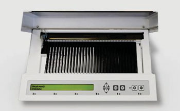
Image: The compact EUROBlotMaster processor for immunoblot strips (Photo courtesy of Euroimmun AG).
Scientists at the Chang Gung University College of Medicine (Taoyuan, Taiwan) measured serum concentrations of four macrophage-mediated factors, including interleukin-6 (IL-6), transforming growth factor-β1 (TGF-β1), vascular cell adhesion protein 1 (VCAM-1) and vascular endothelial growth factor (VEGF), in 23 chronic inflammatory demyelinating polyradiculoneuropathy (CIDP), 28 GBS, 11 Miller-Fisher syndrome (MFS), 40 multiple sclerosis (MS), and 12 Alzheimer's disease (AD) patients, as well as 15 healthy controls.
Serum concentrations of IL-6, TGF-β1, VEGF and VCAM-1 were assessed using enzyme-linked immunosorbent assay (ELISA) (R&D Systems, Minneapolis, MN, USA). Anti-ganglioside (anti-GD1a, anti-GD1b, anti-GM1, anti-GM2, anti- GM3, anti-GQ1b and anti-GT1b) and anti-neuronal antigen (anti- AMPHIPHYSIN, anti-CV2, anti-Hu, anti-Ma2, anti-RECOVERIN, anti-Ri, anti-SOX1, anti-TITIN and anti-Yo) immunoglobulin G (IgG) autoantibodies were measured by using immunoblot strip kit (Euroimmun AG, Luebeck, Germany).
The investigators found that serum TGF-β1 concentration of GBS patients at 35.94 ± 2.55 ng/mL was statistically significantly higher compared with CIDP with a mean of 25.46 ± 1.40 ng/mL, for MFS at 25.32 ± 2.31 ng/mL, MS equaled 21.35 ± 0.90 ng/mL, and AD patients at 22.92 ± 1.82 ng/mL, as well as healthy controls with 23.12 ± 1.67 ng/mL. A positive correlation between serum TGF-β1 concentrations and Hughes' functional grading scales was observed in GBS patients. Serum concentrations of IL-6, VCAM-1 and VEGF were similar between the studied groups. The CSF concentration of TGF-β1 was undetectable by the assay they used.
The authors concluded that that serum concentration of TGF-β1was increased in GBS patients compared to patients with other neurological diseases, including CIDP, MFS, MS and AD, as well as control subjects. These findings implicate TGF-β1 may be involved in the compensatory response to the inflammatory process of GBS. The study will be available in the October 2016 issue of the journal Clinica Chimica Acta.
Related Links:
Chang Gung University College of Medicine
R&D Systems
Euroimmun




Platinum Member

Automated Coagulation Analyzer
Hemolumi H6
Gold Member

Platelet Function Analyzer
PL-12
Gold Member

Blood-Based Protein Biomarker Solution for Alzheimer's Disease
BG-DTi2000.

All-in-One Molecular System
AIO M160