Screening Test Predicts Treatment Response in Metastatic Kidney Cancer
By LabMedica International staff writers
Posted on 19 Oct 2015
Expression levels of a key protein involved in tumor cell survival appear to predict response to standard first-line therapy in patients with metastatic clear cell renal cell carcinoma (mCCRCC).Posted on 19 Oct 2015
Vascular endothelial growth factor pathway (VEGF)-tyrosine kinase inhibitors (TKIs) are essential for the treatment of metastatic renal cell carcinoma patients, but the treatment suffers from a lack of predictive markers.

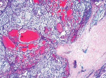
Image: Histopathology of metastatic renal cell carcinoma (Photo courtesy of Dr. Mark R. Wick).
Scientists from Ulsan College of Medicine (Seoul, Republic of Korea) retrospectively studied a total of 91 patients with metastatic renal cell carcinoma (mRCC) between 2006 and 2011. Formalin-fixed, paraffin-embedded tissue samples taken from resected primary tumor at the time of initial diagnosis were exclusively collected; and 1.0-mm-core tissue microarray (TMA) blocks were constructed with two representative cores for each case.
Sections from the TMA blocks were immunostained using the Ventana Benchmark XT automated staining system (Ventana Medical Systems; Tucson, AZ, USA. The number of programmed death ligand-1 (PD-L1) tumor-infiltrating lymphocytes (TILs) were manually counted for each core, and the mean number per unit area (mm2) was calculated for each case. PD-1+ TILs were observed to have increased in the cases with equal to or greater than five TILs per mm2. Tumors with moderate or strong staining in at least 5% of tumor cells were considered positive for PD-L1 expression.
In total, 17.6% of the mCCRCC tumor samples tested positive for PD-L1 expression. The PD-L1-positive tumors were more likely than PD-L1-negative tumors to harbor other aggressive features, including higher tumor grade and sarcomatoid features. The analysis showed that PD-L1 positivity significantly predicted worse treatment response. Only one in eight patients (12.5%) with PD-L1-positive tumors responded to VEGF-TKI therapy, compared with nearly half of patients (46.7%) with PD-L1-negative tumors. Patients with PD-L1-positive tumors also had worse outcomes than those with PD-L1-negative tumors, including worse overall survival and worse progression-free survival.
The authors concluded that PD-L1 expression was significantly related to lack of VEGF-TKI responsiveness and independently associated with shorter survival in mCCRCC patients after VEGF-TKI treatment. PD-L1 may have a predictive and prognostic value for determining the value of VEGF-TKI treatment in patients with mCCRCC. The study was published on September 30, 2015, in the journal the Oncologist.
Related Links:
Ulsan College of Medicine
Ventana Medical Systems


