Antinuclear Antibodies Detected by Automated Immunofluorescence
By LabMedica International staff writers
Posted on 14 May 2013
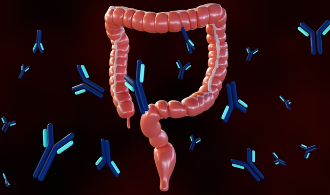
Antinuclear and anticytoplasmic antibodies are important diagnostic markers for systemic rheumatic diseases and autoimmune hepatitis. Posted on 14 May 2013
The diagnostic performance of automated systems for image acquisition and interpretation of indirect immunofluorescence-based tests for antinuclear antibodies are increasingly being used.
Scientists at the Catholic University Leuven (Leuven, Belgium) measured by automated indirect immunofluorescence 268 consecutive samples submitted to the laboratory for antinuclear antibody testing. Included in the study were in 231 patients with a systemic rheumatic disease at the time of diagnosis, 143 blood donors, 134 patients with chronic fatigue syndrome, and 133 diseases controls whose diagnosis of systemic rheumatic disease was excluded.
Antinuclear antibodies were detected by Zenit G-Sight (A. Menarini Diagnostics, Florence, Italy), an automated system for image acquisition and interpretation of indirect immunofluorescence-based tests. The automated fluorescence microscope uses a light-emitting diode (LED) light source with a 450–490 nm bandwidth and is equipped with a motorized precision stage for up to five slides and a Charged Coupled Device (CCD) color camera.
Image acquisition by G-Sight was of high quality and the accuracy of pattern assignment was limited. There was a significant correlation between automated estimation of fluorescence intensity, known as the probability index of positivity and end-point titer. Probability index interval specific likelihood ratios for systemic rheumatic disease increased with increasing level of positivity probability. Probability indexes were higher with the transfected mitotic human epithelioid (Hep-2000) cell substrate (Immunoconcepts; Sacramento, CA, USA) than with the human epithelial cell (Hep-2) substrate. The probability indexes reached a plateau at titer 160 with the Hep-2000 substrate.
The authors concluded that G-Sight offers high quality image acquisition. The accuracy of pattern assignment is limited and needs to be further developed. There was a significant correlation between probability indexes, a measure of fluorescence intensity and antibody titer, and the probability indexes provided by G-Sight, which afforded clinically useful information. Test result specific likelihood ratios help with the interpretation of data generated by automated immunofluorescence analysis of antinuclear antibodies. The study was published on the January 16, 2013, in the journal Clinica Chimica Acta.
Related Links:
Catholic University Leuven
A. Menarini Diagnostic
Immunoconcepts
New
Gold Member
POC Helicobacter Pylori Test Kit
Hepy Urease Test


Electrolyte Analyzer
CBS-4000 (CBS-400)

Manual Pipetting Aid
Pipette Controllers macro