Cell Viability Tests Compared with Microchip Method
By LabMedica International staff writers
Posted on 12 Apr 2011
A technique for the automatic cell viability measurement with a microscopic cell counter and microchip has been evaluated. Posted on 12 Apr 2011
An appraisal has been made of different methods of cell viability testing, an essential tool for performing cell-based studies and clinical laboratory tests.
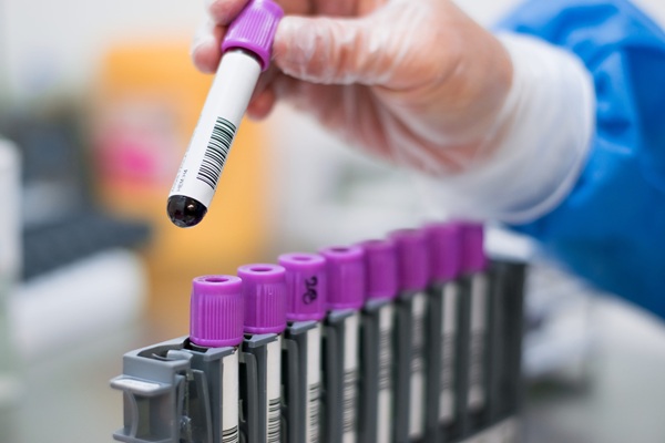
Scientists at Korea University Guro Hospital (Seoul, Korea) compared three different methods to test the viability of blood cells. Blood was drawn from 11 healthy volunteers and mononuclear cells were separated immediately from the heparinized whole blood, and the viable cells were diluted subsequently down from 100% to, 75%, 50%, 25%, and 1%. The cell viability tests were performed simultaneously with the following three methods: the conventional manual trypan blue exclusion method; the flow cytometry measurement with propidium iodide stain; and the newly developed automated fluorescence microscopic cell counter with microchip.
The linearity, precision, and correlations from three methods were analyzed and compared. The correlations data from the microscopic cell counter were in good agreement with both the conventional trypan blue method and the flow cytometry. The precision and linearity from the microscopic cell counter method with microchip were superior in comparison with the conventional method. The new microscopic cell counter with microchip, known as Adam (NanoenTek, Inc.; Seoul, Korea), was further developed and improved to produce the results within five minutes, including all procedural steps.
In the manual trypan blue stain, only 200 cells were counted to measure their viability, but in Adam, larger number of cells, approximately between 1,000 and 3,000 cells, were counted. Greater number of counted cells and the repetitions of the experiments by the microscopic cell counter and microchip for the analyses provided the better statistical significance. The real-time cellular images and archived data could also be used for analyzing other parameters. The study was published online on March 15, 2011, in the Journal of Clinical Laboratory Analysis.
Related Links:
Korea University Guro Hospital
NanoenTek, Inc.
 Assay.jpg)
Gold Member
H-FABP Assay
Heart-Type Fatty Acid-Binding Protein Assay


New
Gold Member
Pre- Eclampsia Control
Acusera Pre-Eclampsia Control
New
Fully-auto Specific Protein (Nephelometry) Analyzer
PA240