Peripheral Blood Smears Still Need Evaluation
By LabMedica International staff writers
Posted on 22 Jun 2017
When the first automated hematology analyzers appeared in clinical laboratories in the 1960s, they ushered in a welcomed workflow change for bench technologists. These automated analyzers replaced hemocytometers, though the need for differential counting remained.Posted on 22 Jun 2017
This evolution in hematology workflows has continued to this day, with automated instruments performing ever more cellular analysis, resulting in more focused roles for technologists and pathologists. However, certain characteristics of peripheral blood morphology still do not lend themselves easily to evaluation by automated analyzers.
.")
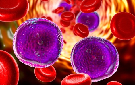
Image: A white blood cell among red blood cells (Photo courtesy of HealthTap).
A clinical associate professor at the University of Florida (Gainesville, FL, USA) has written that one limitation that has remained constant from the earliest hematology analyzers to today’s cutting-edge flow cytometers is that a single cell still must pass through an aperture for analysis. In order to maintain laminar flow, the cell must also be sphered, which is most often accomplished with a proprietary sphering reagent. The exact classification of abnormally shaped red cells, for example, sickle cells, target cells, and schistocytes, still requires morphologic review of stained slides.
In addition red cell and white cell inclusions, particularly infectious organisms such as malaria or histoplasmosis, can be seen in stained blood smears but are not routinely detected by most automated hematology analyzers. Because of the extensive morphologic variability of many circulating hematologic malignancies, automated systems cannot precisely characterize these cells. Most analyzers, however, aid in characterizing these cells by pre-classifying them as abnormal (through large unstained cell classification or flagging) and prompting manual review of slides.
Analyzers that have digital morphology capabilities, such as the CellaVision or the Bloodhound systems, are inaugurating a new era of cellular analysis. As these instruments’ algorithms continue to be refined, this technology might evolve from a pre-classifier method to a more enhanced and robust method for precise characterization. The accuracy of an automated differential count depends on the analytical system used. However, given that most automated counters literally characterize thousands of white cells for each analysis, the classic 100-cell manual differential count in comparison falls short when it comes to precision. Sherri D. Flax, MD, published her article on June 1, 2017, in the journal Clinical Laboratory News.
Related Links:
University of Florida
.jpg)
Gold Member
Clinical Chemistry Assay
Sorbitol Dehydrogenase (SDH)


Immunofluorescence Analyzer
IFA System

Electrolyte Analyzer
BKE-B