Optical Microscope Can Image Flow of Individual Blood Cells
By LabMedica International staff writers
Posted on 31 May 2012
A new device uses a technique called spectrally encoded confocal microscopy (SECM), which creates images by splitting a light beam into its constituent colors.Posted on 31 May 2012
The rainbow of light has been used to image the flow of individual blood cells providing a noninvasive test, which promises rapid, pain free diagnoses. The optical instrument is able to provide high-resolution images of blood coursing through the veins without the need for harsh and short-lived fluorescent dyes.
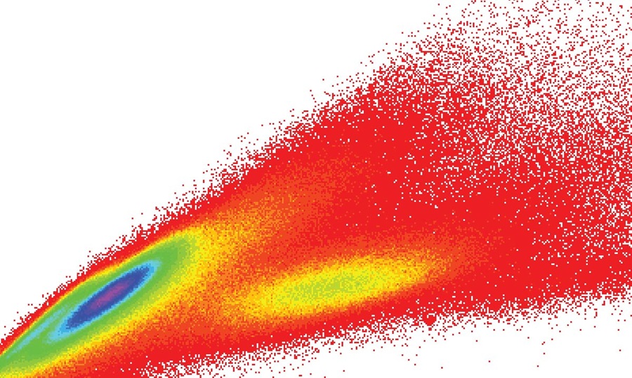
 A single line within a blood vessel is imaged with multiple colors of light that encode lateral positions. (b) A single cell crossing the spectral line produces a two-dimensional image with one axis encoded by wavelength and the other by time (Photo courtesy of the Technion, Israel Institute of Technology, Biomedical Optics Lab).")
Image: Image acquisition in SEFC. (a) A single line within a blood vessel is imaged with multiple colors of light that encode lateral positions. (b) A single cell crossing the spectral line produces a two-dimensional image with one axis encoded by wavelength and the other by time (Photo courtesy of the Technion, Israel Institute of Technology, Biomedical Optics Lab).
.")
Image: In vivo imaging in microvessels. Extracting the fractional area occupied by RBCs in a vessel using manual segmentation for assessing hematocrit levels. Red regions correspond to areas occupied by RBCs (Photo courtesy of the Technion, Israel Institute of Technology, Biomedical Optics Lab).
The colors are spread out in a line from red to violet. To scan blood cells in motion, a probe is pressed against the skin of a patient and the rainbow-like line of light is directed across a blood vessel near the surface of the skin. As blood cells cross the line, they scatter light, which is collected and analyzed. The color of the scattered light carries spatial information, and computer programs interpret the signal over time to create 2-D images of the blood cells.
Using the new microscope, scientists imaged the blood flowing through a vessel in the lower lip of a volunteer. They successfully measured the average diameter of the red and white blood cells and calculated the percent volume of the different cell types, a key measurement for many medical diagnoses.
Lior Golan, a graduate student in the biomedical engineering department at the Israel Institute of Technology, or Technion (Haifa, Israel), noted that by eliminating the long waiting time for blood test results, "the new microscope might help spotlight warning signs, like high white blood cell count, before a patient develops severe medical problems. The portability of the device could also enable doctors in rural areas without easy access to medical labs to screen large populations for common blood disorders."
The narrow field of view of the microscope made it difficult for the team to locate suitable capillary vessels to image. Therefore, the scientists added a green light-emitting diode (LED) and camera to the system to provide a wider view in which the blood vessels appeared dark because hemoglobin absorbs green light. “Unfortunately, the green channel does not help in finding the depth of the blood vessel,” noted Dr. Golan. “Adjusting the imaging depth of the probe for imaging a small capillary is still a challenge we will address in future research.”
The scientists are also working on a second-generation system with higher penetration depth. The new system might expand the range of possible imaging sites beyond the inside lip, which was selected as a test site because it is rich in blood vessels, has no pigment to block light, and doesn’t lose blood flow in trauma patients.
Additional steps include work to miniaturize the system for ease of transport and use. “Currently, the probe is a bench-top laboratory version about the size of a small shoebox,” said Dr. Golan. “We hope to have a thumb-size prototype within the next year.”
Other blood-scanning systems with cellular resolution do exist, but they are far less practical, relying on bulky equipment or potentially harmful fluorescent dyes that must be injected into the bloodstream.
A paper describing the device will appear on June 1, 2012, in the Optical Society's (OSA) open-access journal Biomedical Optics Express.
Related Links:
Technion
Technion Biomedical Optics Lab

Gold Member
Quantitative POC Immunoassay Analyzer
EASY READER+


HPV Test
Allplex HPV28 Detection

Multi-Chamber Washer-Disinfector
WD 390