Diffraction Phase Microscopy Reveals Mechanics of Erythrocyte Membranes
By LabMedica International staff writers
Posted on 29 Apr 2010
A novel measurement technique called diffraction phase microscopy uses two beams of light in contrast to other microscopes that only use one.Posted on 29 Apr 2010
Using diffraction phase microscopy a team of scientists developed a model that could lead to breakthroughs in screening and treatment of blood-cell-morphology diseases, such as malaria, sickle-cell disease, and spherocytosis. It could also be used to screen banked blood for membrane flexibility before transfusion, since stored blood often undergoes cellular shape changes.
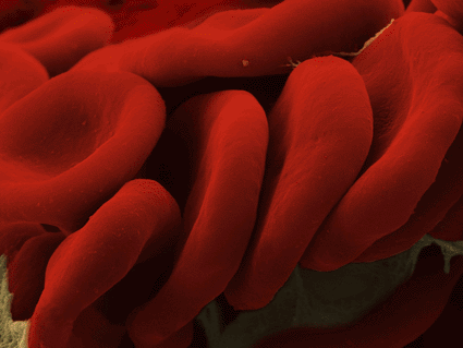
Image: Erythrocytes (photo courtesy Mustafa Mir, Sam Copeland, and Gabriel Popescu. Additional information may be found at http://light.ece.uiuc.edu).
In circulation, erythrocytes or red blood cells (RBCs) must contort to squeeze through capillaries half their diameter. Their flexibility and resilience come from their membrane structure, which couples a typical lipid bilayer with an underlying matrix of protein. However, knowledge of the membrane's mechanics is very limited.
Prof. Gabriel Popescu of the electrical and computer engineering department at the University of Illinois at Urbana Champaign (Champaign, IL, USA) and colleagues were able to see nanoscale membrane fluctuations in live cells, and to measure them quantitatively. The group published its findings in the April 13, 2010 publication of the Proceedings of the [U.S.] National Academy of Sciences (PNAS).
In addition to normal cells, the team also measured two other morphologies: bumpy RBCs called echinocytes and round ones called spherocytes. They discovered that these deformed cells display less flexibility in their membranes, a finding that could provide insight into mechanics and treatment of diseases.
Because diffraction phase microscopy measures live cells without physically manipulating or damaging them, it also could be used to evaluate medications being developed to treat blood cell morphology diseases, according to Prof. Popescu. "We can study the mechanics of a single cell under different pharmacological conditions, and I think that would be ideal for testing drugs," he said.
Related Links:
University of Illinois at Urbana Champaign




Platinum Member

Automated Coagulation Analyzer
Hemolumi H6


Gold Member

Platelet Function Analyzer
PL-12

Gold Member

Flocked Fiber Swabs
Puritan® Patented HydraFlock®