Cutaneous Leishmaniasis Species Identified by Mass Spectrometry
By LabMedica International staff writers
Posted on 18 Jun 2014
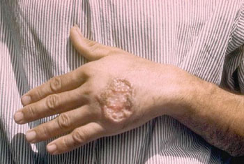
Cutaneous leishmaniasis (CL) is caused by several Leishmania species that are associated with variable outcomes before and after therapy and optimal treatment decision is based on an accurate identification of the infecting species.Posted on 18 Jun 2014
Current methods to type Leishmania isolates are relatively complex and slow and therefore, the initial treatment decision is generally presumptive, the infecting species being suspected on epidemiological and clinical grounds. A simple method to type cultured isolates would facilitate disease management.
Scientists at the University Pierre et Marie Curie (Paris, France) performed parasitological diagnosis and analysis by lesion scraping, biopsy or aspirate followed by direct examination of Giemsa-stained smears, histological analysis, Giemsa-stained tissue sections, culture or polymerase chain reaction (PCR). They also analyzed promastigote pellets from 46 strains cultured in monophasic medium, including 20 short-term cultured isolates from French travelers, 19 with CL, and one with visceral leishmaniasis and clinical isolates were analyzed in parallel with Multilocus Sequence Typing (MLST).
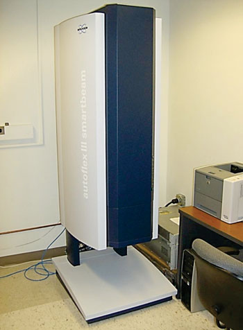
The promastigote pellets were analyzed by Matrix Assisted Laser Desorption Ionization Time-of-Flight (MALDI-TOF) technology and performed on a BrukerAutoflex I MALDI-TOF mass spectrometer (Bruker-Daltonics, Bremen, Germany). Automatic dendrogram analysis generated a classification of isolates consistent with reference determination of species based on MLST or 70 kDa heat shock protein gene (hsp70) sequencing. An analysis of the spectra, based on a very simple, database-independent analysis of spectra, showed that the mutually exclusive presence of two pairs of peaks discriminated isolates considered by reference methods to belong either to the Viannia or Leishmania subgenus, and that within each subgenus presence or absence of a few peaks allowed discrimination to species complexes level.
The authors concluded that analysis of cultured Leishmania isolates using mass spectrometry allows a rapid and simple classification to the species complex level consistent with reference methods, a potentially useful method to guide treatment decision in patients with cutaneous leishmaniasis. The intuitive interpretation of spectra was well-suited for allowing for close interactions between parasitologists and clinicians. The study was published on June 5, 2014, in the journal Public Library of Science Neglected Tropical Diseases.
Related Links:
University Pierre et Marie Curie
Bruker-Daltonics


