Simplified Microchip Electrophoresis Rapidly Separates Urinary Proteins
By LabMedica International staff writers
Posted on 27 Mar 2014
A urine protein test is to detect the presence of protein in a person's urine, while in healthy individuals, there is only very low concentration of protein in urine, but in renal patients, the concentration of urine protein will increase considerably.Posted on 27 Mar 2014
Urine protein test has been widely used in clinics, but to determine the type of proteinuria is usually difficult due to technical limitations. A rapid and simple method to separate and determine urine proteins by a microchip electrophoresis (ME) system has been developed which can be completed rapidly.
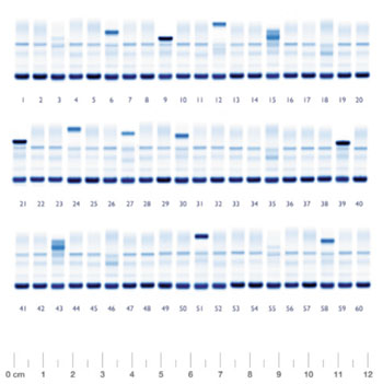
Image: Conventional Agarose Gel Electrophoresis for proteins (Photo courtesy of Helena).
Scientists at the Affiliated Hospital of Nantong University (Jiangsu Province, China) developed quick and straightforward method to separate urine proteins with ME. By testing different experimental conditions, the optimal parameters were established and the duration of the analytical run takes less than four minutes. They compared ME and conventional agarose gel electrophoresis (AGE) and by analyzing 248 urine samples.
The chip electrophoresis apparatus consisted of an optical module, a control module, and a detection module (Shanghai Institute of Microsystem and Information Technology; China). The 214 nm UV light was obtained by a hologram grating and focused the separation channel to detect the presence of protein. The light absorbed by a sample was gathered by a photomultiplier tube (Hamamatsu; Beijing, China) and converted to electric signals, which were then processed by a filtering and amplifying circuit and input to a computer.
The patient group contained 178 urine samples, of which no protein peak was detected for 7 samples, 60 were selective proteinuria with primary protein of albumin, 105 were nonselective proteinuria with various molecular weight proteins, and 6 were overflow proteinuria. The same results were obtained by conventional AGE (Helena; Beaumont, TX, USA) as the ME.
The authors concluded that the assay was rapid, simple, and convenient, with electrophoretic time only four minutes with good reproducibility. The electrophoresis apparatus and detection system were cheap and microfluidic chip could be repeated. The newly established ME provided a useful clinical tool with broad potential applications for the diagnosis, treatment, and prognosis of renal and related diseases. The study was published in the March 2014 issue of the Journal of Clinical Laboratory Analysis.
Affiliated Hospital of Nantong University
Shanghai Institute of Microsystem and Information Technology
Helena
Related Links:




Platinum Member

Automated Coagulation Analyzer
Hemolumi H6
Gold Member

Quantitative POC Immunoassay Analyzer
EASY READER+
New

Slide Scanner System
NanoZoomer S540MD
New

Portable POCT Blood Gas Analyzer
BD100