Mass Cytometry Profiles to Aid Drug Development
By LabMedica International staff writers
Posted on 15 May 2017
Cancer researchers used a novel mass cytometry technique to reveal potential biomarkers for detection of renal cell carcinoma and to identify targets for development of immunotherapeutic agents to treat this essentially incurable disease.Posted on 15 May 2017
Renal cell carcinoma (ccRCC) is a common and lethal uro-genital cancer. Around 50% of ccRCC patients develop metastases despite treatment, and less than 10% of these patients survive five years. Targeted treatments such as tyrosine kinase inhibitors do not cure the disease, but novel checkpoint immunotherapies have shown considerable promise in some patients. Thus, new avenues to guide immunotherapy-based treatments are urgently needed.
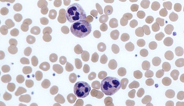
 and T-cells (blue) present in the microenvironment of renal cell carcinoma (Photo courtesy of Karina Silina, University of Zurich).")
Image: Fluorescent imaging of a tumor section identifies different types of macrophages (green) and T-cells (blue) present in the microenvironment of renal cell carcinoma (Photo courtesy of Karina Silina, University of Zurich).
Towards this end, investigators at the University of Zurich used mass cytometry with extensive antibody panels to perform in-depth immune profiling of samples from 73 ccRCC patients and five healthy controls. Mass cytometry enabled the quantification of more than 50 readouts at the single-cell level by combining metal isotope-labeled antibodies with mass spectrometry detection. With its ability to analyze millions of cells in a short time at low cost, mass cytometry represents the method of choice to assess the phenotypic diversity of T-cells and tumor-associated macrophages (TAMs) present in the tumor microenvironment in large patient cohorts.
The investigators reported in the May 4, 2017, issue of the journal Cell that by screening 3.5 million cells, they had identified 17 tumor-associated macrophage phenotypes and 22 T-cell phenotypes. This data expanded the view of T-cell immunosuppression phenotypes, suggested links between a TAM phenotype and populations of regulatory T-cells and CD8+ immunosuppressed T-cells, and identified an immune cell composition within the tumor microenvironment that was correlated with progression-free survival.
"The previous picture of immune defense was correct, but coarse," said first author, Dr. Stéphane Chevrier, a researcher in the institute of molecular life sciences at the University of Zurich. "With our methods to analyze individual immune cells, we have been able to create an immunological atlas of the tumor environment for the first time with high resolution and in a large patient cohort. As a result, many more facets have now come to light."

Gold Member
Flocked Fiber Swabs
Puritan® Patented HydraFlock®


New
Urine Analyzer
respons® UDS100

Rapid Sepsis Test
SeptiCyte RAPID