Nanoscale Gene "Ignition Switch” May Help Target and Treat Cancer
By LabMedica International staff writers
Posted on 13 Jan 2011
A proof of principal study using laboratory mice has shown that a set of genetic instructions encased in a nanoparticle can be used as an "ignition switch” to rev up gene activity that aids cancer detection and treatment. Posted on 13 Jan 2011
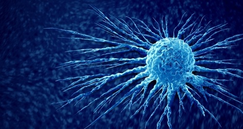
The switch, called a promoter, is a set of chemical letters that interacts with DNA to turn on gene activity. In this case, the scientists, at Johns Hopkins University (Baltimore, MD, USA) and the Virginia Commonwealth University (VCU; Richmond, USA), used a promoter called PEG-Prom (promoter of progression elevated gene-3), cloned by VCU researcher Paul Fisher, PhD PEG-Prom is activated only when inside cancer cells, not in normal ones.
"With current imaging devices like CT [computed tomography] and PET [positron emission tomography], we can tell if something is wrong in a patient, but we don't have definitive tools to distinguish cancer from inflammation or infection,” said Martin Pomper, MD, PhD, professor of radiology at Johns Hopkins. "It generally takes at least one month after giving patients certain cancer treatments before existing imaging tools can measure the patient's response to the therapy.”
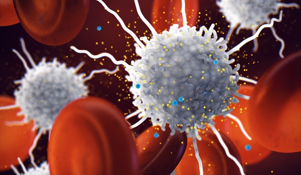
To distinguish cancer cells from normal cells, Johns Hopkins scientists connected PEG-Prom to either a gene that produces firefly luciferase or a gene called HSV1tk, which initiates a chemical reaction with radioactive labels inside the cell that can be detected by imaging devices. Once inside a cancer cell, the PEG-Prom switch is turned on, and it activates either the luciferase or HSV1tk gene. Then, the scientists packed the PEG-Prom/gene combination into tiny spheres --approximately 50,000 times smaller than the head of a pin--and intravenously injected the nanoparticles into mice with either metastatic breast cancer or melanoma.
The study's findings, published in the December 12, 2010, online edition of the journal Nature Medicine, revealed a 30-fold difference in identifying cancer cells containing luciferase and normal cells that did not contain the substance. Similar findings were seen in cancer cells filled with the radioactive labels and normal ones that were not.
Dr. Pomper reported that the technique could likely be used in any cancer, and the nanoparticle and HSV1tk gene used in the current study have been evaluated earlier in clinical studies unrelated to his studies. In addition to diagnostic and monitoring tools, the technique could be designed to deliver therapies to the heart of cancer cells. One approach, he noted, is to utilize radioactive isotopes to make cancer cells radioactive from the inside, instead of delivering radiation to the patient externally.
Nevertheless, according to Dr. Pomper, such a technique would be restricted to identifying tumors that are 2 mm or larger, amounting to millions of cells, because current imaging devices cannot detect anything smaller. He also noted that certain doses of nanoparticles could be toxic, so his team is conducting tests to find the best nanoparticle.
Related Links:
Johns Hopkins University
Virginia Commonwealth University

New
Gold Member
Automatic Hematology Analyzer
CF9600


New
Creatinine/eGFR Meter
StatSensor® Creatinine/eGFR Meter

New
Japanese Encephalitis Test
Japanese Encephalitis Virus Real Time PCR Kit