Atomic Force Microscopy Differentiates Cancerous from Normal Cells
By LabMedica International staff writers
Posted on 29 Apr 2009
Using an atomic force microscope, scientists discovered that cancer cells and normal cells had different cell surface properties. The difference in the brush layers covering the cell surface might have a biological significance, and could be used for detection of cancer.Posted on 29 Apr 2009
Brushes on the cell surface, mostly consisting of microridges and tiny hairs called microvilli, are important for interacting with the external environment. The investigators processed force measurements--taken from the cell surface using an atomic force microscope--according to a model that accounts for these brushes, to quantitatively show that cancerous cells are different. Normal cells have brushes of one length, whereas cancerous cells have mostly two brush lengths of significantly different densities.
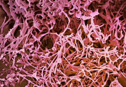
Image: Colored scanning electron micrograph (SEM) of the surface of a cervical cancer cell, showing microvilli strands that are small projections of the cell’s cytoplasm (Photo courtesy of Steve Gschmeissner / SPL).
The scientific group was led by Igor Sokolov, director of the Nanoengineering and Biotechnology Laboratories Center at Clarkson University (Potsdam, NY, USA). "Cancer cells are traditionally detected by biochemical means," said Professor Sokolov. "However, despite many years of success, those methods have not resulted in defeating cancer. Therefore, it is very important to search for alternative, nontraditional ways of looking at cancer. Investigation of the physical properties of cell surfaces might be one such nontraditional way."
The authors also demonstrated that the difference found in cell surface "brush" is practically impossible to find with other microscopic methods.
The study appeared online in Nature Nanotechnology, on April 13, 2009.
Related Links:
Clarkson University




Platinum Member

Automated Coagulation Analyzer
Hemolumi H6
Gold Member

Neonatal Heel Incision Device
Tenderfoot
New

Portable POCT Blood Gas Analyzer
BD100
New

Gastrointestinal Panel
Xpert® GI Panel