AI-Powered Atlas Maps Immune Structures Linked to Cancer Outcomes
Posted on 29 May 2026
Tertiary lymphoid structures are emerging as important indicators of antitumor immunity, but their heterogeneity and spatial context within tumors remain difficult to capture through routine diagnostics. Scalable tools are also needed to translate these features into reproducible biomarkers for prognosis and therapy response. New findings demonstrate an artificial intelligence approach that builds a pan-cancer atlas of these structures and derives a composite score linked to patient outcomes.
The University of Texas MD Anderson Cancer Center (Houston, TX, USA) developed scalable AI frameworks and a pan-cancer spatial atlas to characterize tertiary lymphoid structures (TLSs) across multiple tumor types. The work, published in Science, indicates that TLS maturation state, spatial location, and cellular composition within tumors carry clinically meaningful information beyond simple presence or absence. The team also introduced a TLS composition score intended to stratify patients by prognosis and treatment response.
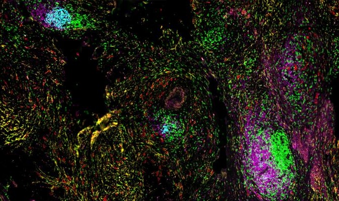
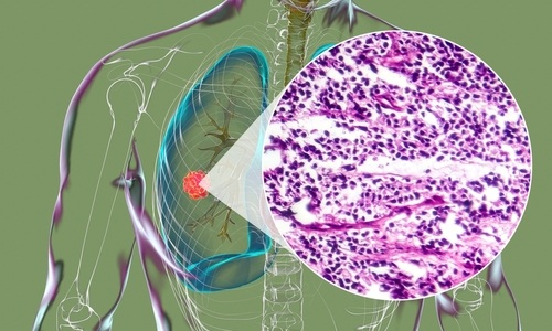
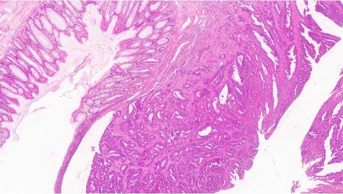
The atlas was constructed by detecting, profiling, and classifying TLSs from spatial omics datasets and routine pathology slides. Using these frameworks, investigators analyzed 340 samples spanning 12 cancer types to define TLS features in their native spatial context and to identify transcriptional programs associated with TLS maturation. The study reports that as TLSs mature, they exhibit greater organization with coordinated shifts in immune, stromal, and vascular components, and that proximity to tumor cells aligns with spatial gradients of tumor signaling.
To scale translation to clinical workflows, the AI model rapidly identified and classified TLSs from whole-slide pathology images. Across 10 independent cohorts encompassing more than 3,000 whole-slide images and 25,088 TLSs, the derived composition score integrated TLS number and maturation states. This approach outperformed conventional TLS measures for stratifying prognosis and treatment response across diverse cancers and treatment contexts, according to the report.
The authors note that many TLSs remain immature and are sometimes located away from tumor regions, highlighting questions about how to promote maturation and enhance spatial interactions with tumor cells. They state that the composite scoring method requires validation in prospective clinical trials and, if successful, could support broader use of TLS profiling with routinely generated pathology images.
“Prior to this study, most of the focus on TLSs as biomarkers was simply on whether or not they were present, and—in some cases—whether they were mature. Here, we show that we can go much deeper. TLSs in tumor tissues are much more complex than that. Their maturation state, spatial location and composition within tumors can tell us critical information about the tumor immune microenvironment, treatment response and clinical outcomes,” said Linghua Wang, M.D., Ph.D., professor of Genomic Medicine and executive director and head of the Center for Cellular Language Intelligence at The University of Texas MD Anderson Cancer Center.
Related Links
UT MD Anderson Cancer Center