Changes in Eye Tissue May Enable Early Detection of Brain Diseases
By LabMedica International staff writers
Posted on 11 Oct 2016
Research with mouse models has shown that at least some diseases of the central nervous system (CNS) manifest as pathological changes in the retina of the eye and that these changes may be detected earlier than brain changes. The findings suggest that eye examination could be used for minimally invasive screening for these diseases.Posted on 11 Oct 2016
Retina tissue can be considered an integral part of the central nervous system (CNS). During fetal development, it matures from part of the brain and its innervation closely resembles that of the brain. Retinal structure and function can be readily examined with noninvasive or minimally invasive methods, whereas direct brain examination has numerous limitations. If, at least for some brain diseases, the health status of the brain could be indirectly assessed through the eyes, diagnostic screening could become more efficient.
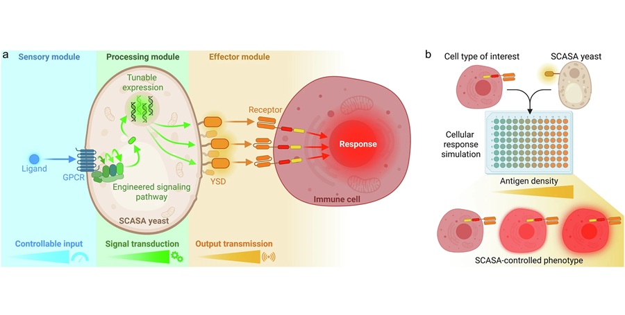
; Red: mutant huntingtin protein (mHtt). (A) A low magnification picture illustrates GFAP-ir astrocytes and mHtt deposits from the retinal wholemount of 12-week-old R6/2 (Huntington’s disease model) mouse. Scale bar = 50 µm. (B) A detailed confocal analysis of GFAP positivity, mHtt immunoreactivity, and DAPI counterstain (blue) revealed no colocalization of GFAP and mHtt. Scale bar = 20 µm (Image courtesy of PLoS One).")
Image: An experiment examining retina tissue for mHtt deposition in GFAP-ir astrocytes in R6/2 mouse model of Huntington’s Disease. Green: glial fibrillary acidic protein (GFAP); Red: mutant huntingtin protein (mHtt). (A) A low magnification picture illustrates GFAP-ir astrocytes and mHtt deposits from the retinal wholemount of 12-week-old R6/2 (Huntington’s disease model) mouse. Scale bar = 50 µm. (B) A detailed confocal analysis of GFAP positivity, mHtt immunoreactivity, and DAPI counterstain (blue) revealed no colocalization of GFAP and mHtt. Scale bar = 20 µm (Image courtesy of PLoS One).
In his PhD project at the University of Eastern Finland at Kuopio (Kuopio, Finland), Dr. Henri Leinonen and colleagues investigated functional abnormalities of the retina using mouse models of human CNS diseases. Electroretinography (ERG) and visual evoked potentials (VEP) were chosen as research techniques, since similar methodology can be applied in both laboratory animals and humans. ERG can precisely track the function of retina using corneal or skin electrodes, whereas VEP measures the function of visual cortex.
These methods were used to test different attributes of vision in 3 distinct genetically engineered mouse models of human CNS diseases. Also, basic life science methods were used to test the correlation between functional abnormalities and the anatomical status of the retina.
Day and color vision -associated retinal dysfunction was found in a mouse model of Huntington´s disease (HD) while the mouse was presymptomatic. Retinal structure remained relatively normal, even in an advanced disease state, although aggregation of toxic mutated huntingtin-protein was widespread in the diseased mouse retina. Although the retinopathy in mice is exaggerated compared to human HD patients, the finding is partly in line with patient data showing impaired color vision but no clear-cut anatomical retinopathy.
In a mouse model of Alzheimer´s disease (AD), the researchers observed abnormality in night vision -associated retinal function. Specifically, rod-mediated inner retinal responses to dim light flashes were faster in diseased mice than in their wild-type controls. The observation may be explained by impaired cholinergic neurotransmission that is also partly causative for the deterioration of memory in AD.
In a mouse model of late infantile neuronal ceroid lipofuscinosis (NCL), a pediatric neurological disease, the researchers described retinal degenerative changes that mimic the characteristic pathology of age-related macular degeneration (AMD). These included impaired function of retinal pigment epithelium and subsequent blindness due to photoreceptor atrophy and death. It has been postulated that the retinal degeneration in human patients progresses similarly.
Adding to the growing body of evidence, the results showed that functional changes of the retina occur in mouse models of three human CNS diseases whose phenotype, age of onset, and pathological mechanism clearly differ from each other. Visual impairment was the fastest progressive symptom in two models tested.
The findings support the idea of eye examinations as potential screening tools for CNS diseases. Development of efficient, safe, and economic screening is imperative since the diagnosis of these diseases is often obtained only in the advanced disease state, when as such satisfactory remedies are poorly effective. Since eye and vision research can be conducted noninvasively, advancement of trials from the preclinical to the clinical phase could be relatively fast.
Dr. Leinonen’s doctoral dissertation, entitled “Electrophysiology of visual pathways as a screening tool for neurodegenerative diseases: evidence from mouse disease models”, is available for download. The findings were published in PLoS One, the Journal of Alzheimer’s Disease, and most recently in the journal Human Molecular Genetics.
Related Links:
University of Eastern Finland

Gold Member
Hybrid Pipette
SWITCH


Gel Cards
DG Gel Cards
Automatic Hematology Analyzer
DH-800 Series