Automated Test Strongly Predicts Ovarian Cancer Survival
By LabMedica International staff writers
Posted on 06 Oct 2016
The tumor microenvironment is pivotal in influencing cancer progression and metastasis and different cells co-exist with high spatial diversity within a patient, yet their combinatorial effects are poorly understood.Posted on 06 Oct 2016
Ovarian cancer is the most fatal gynecological malignancy and the vast majority of ovarian cancer deaths are from high-grade serous carcinoma (HGSOC) with hardly any change in overall survival in the past years. This is partly due to the distinctive biology of HGSOC: the lack of an anatomical barrier in the peritoneal cavity to cancer cell dissemination likely originated from the fallopian tube or precursor cells in the ovary.

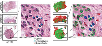
Image: A histology image analysis and validation of the automatic test (Photo courtesy of the Institute of Cancer Research).
Scientists at the Institute of Cancer Research (London, UK) and their colleagues assessed the cell 'ecosystems' at sites where ovarian cancer has spread round the body strongly predicts the chances of surviving from the disease. A total of 61 patients with International Federation of Gynecology and Obstetrics (FIGO) stage III-IV HGSOC ovarian cancer with at least one locally advanced resectable metastasis site were identified. A total of 192 paraffin embedded blocks from seven local metastasis sites (61 ovaries, 51 omentum, 48 peritoneum, nine appendixes, 20 lymph nodes, one spleen, two umbilicus) were obtained from 61 patients.
Histological sections of multiple metastases from paraffin-embedded tumor blocks were generated, digitalized using the Aperio system, (Leica Biosystems) and analyzed using open source R package CRImage. Cells were classified based on their morphological differences of the nucleus positive for hematoxylin stain without using immunohistochemical target stains. Immune cells typically display small, round and homogeneously basophilic nuclei; cancer cells in general have nuclei of larger size and greater variability in texture and shape.
The team found a difference in survival rates between women with high and low levels of diversity at these metastatic sites. The fully automated test could identify those women who have the most life-threatening disease, and who urgently need the most aggressive treatment. The test gives a score for metastasis diversity, known as MetDiv, based on whether a patient's sites of cancer spread have one dominant cell type (low score) or a more diverse cell population containing immune or connective tissue cells (high score). Survival was far poorer among women with high diversity scores than those with low scores. Just 9% of women with diverse metastases survived five years from diagnosis, compared with 42% of those whose metastases were dominated by one cell type. A high diversity score was a stronger predictor of poor survival than any of the clinical factors currently used to try to assess a woman's prognosis.
Yinyin Yuan, PhD, Team Leader in Computational Pathology and co-author of the study, said, “We have developed a new test to assess the diversity of metastatic sites, and use it to predict a woman's chances of surviving their disease. More work is needed to refine our test and move it into the clinic, but in future it could be used to identify women with especially aggressive ovarian cancers, so they can be treated with the best possible therapies available on the NHS or through clinical trials.” The study was published on September, 19, 2016, in the journal Oncotarget.
Related Links:
Institute of Cancer Research


